
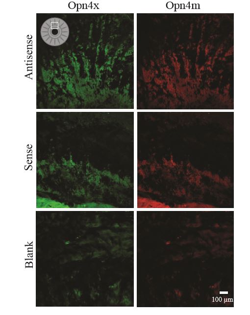
- The mRNA coding for Opn4x and Opn4m are both found in the iris of the turtle (Fig 1).
- This conclusion is supported by the calibration of signals (Fig 3) and the quantitative analysis that was performed, comparing the signals from the
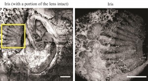
Figure 1
antisense slides to those of the sense and blank slides
- The qPCR studies that were performed in Dr. Dearworth’s lab also support this finding (Dearworth et al., 2011; Goldberg, 2012; Dearworth et al., 2012)
- This result is consistent with prior studies performed on the Xenopus and the chicken, which are also non-mammals (Provencio et al., 1998; Chaurasia et al., 2005; Bellingham et al., 2006)
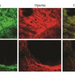
Figure 2
- This conclusion is supported by the calibration of signals (Fig 3) and the quantitative analysis that was performed, comparing the signals from the
- The dim fluorescence found in the blank and sense iris slides is likely caused by autofluorescence
- It is possible that the turtle iris has fluorescent pigments like the irises of birds (Tilloston & Oliphant, 1990)
- The likelihood of the presence of fluorescent pigments in the turtle iris is also increased by the fluorescent oil droplets that turtles and birds share (Ohtsuka, 1985)
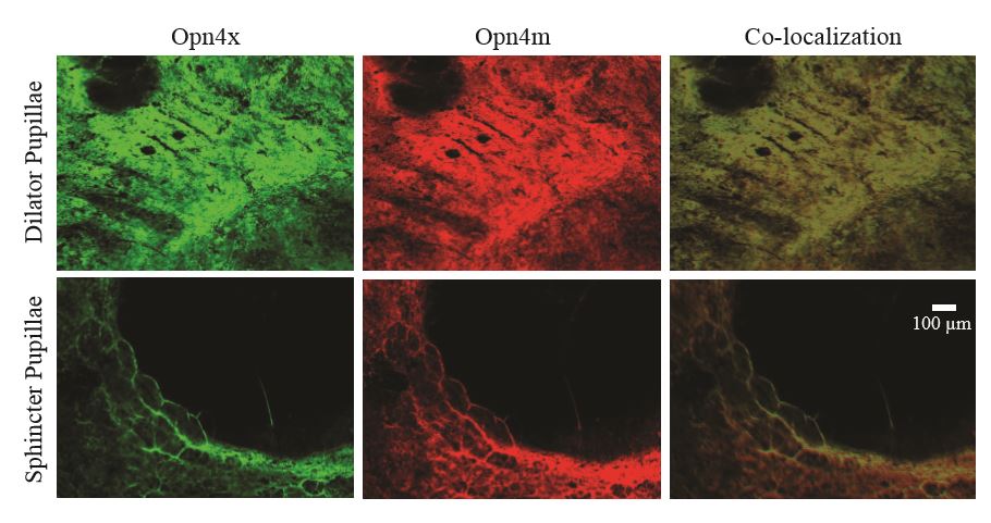
- Both the Opn4x and Opn4m melanopsin isoforms appear to be found within the iris, sphincter, and dilator of turtle eyes, and they also appear to overlap
- This conclusion cannot be supported by the images produced by this study because it is not possible to distinguish the different types of muscle cells in those images
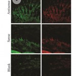
Figure 3
- This conclusion cannot be supported by the images produced by this study because it is not possible to distinguish the different types of muscle cells in those images
- It may be possible to identify the cell types that express the melanopsin isoforms by using a preassembled genome of the red-eared slider turtle, GenBank SRX217618, and then purchasing the antibodies necessary to carryout immunohistochemistry (IHC)
- Using IHC experiments, the specific locations of the cells could then be identified, if probes are created that label for actin and Opn4x and Opn4m proteins
- By creating probes that label for actin, the type of muscle (smooth or skeletal) could potentially be distinguished, for they have difference muscle organizations
- Using IHC experiments, the specific locations of the cells could then be identified, if probes are created that label for actin and Opn4x and Opn4m proteins
- By identifying which cell types express the melanopsin isoforms, we could then possibly find an explanation for the slow pupillary light response that turtles demonstrate
- Although the presence of the melanopsin mRNA does not say for sure that the melanopsin isoforms are expressed, the evidence still supports this possibility

http://www.rockland-inc.com/ihc-products.aspx